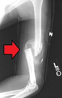
Fibrocartilage callus
Appearance
This article needs more reliable medical references for verification or relies too heavily on primary sources. (June 2019) |  |

A fibrocartilage callus is a temporary formation of fibroblasts and chondroblasts which forms at the area of a bone fracture as the bone attempts to heal itself. The cells eventually dissipate and become dormant, lying in the resulting extracellular matrix that is the new bone. The callus is the first sign of union visible on x-rays, usually 3 weeks after the fracture. Callus formation is slower in adults than in children, and in cortical bones than in cancellous bones.[1]
See also
[edit]References
[edit]- ^ Mirhadi, Sara; Ashwood, Neil; Karagkevrekis, Babis (2013). "Factors influencing fracture healing". Trauma. 15 (2): 140–155. CiteSeerX 10.1.1.834.3328. doi:10.1177/1460408613486571. S2CID 73325632.
- Morgan, Elise F., et al. “Overview of Skeletal Repair (Fracture Healing and Its Assessment).” Methods in Molecular Biology Skeletal Development and Repair, 2014, pp. 13–31. doi:10.1007/978-1-62703-989-5_2
External links
[edit]- Bony+callus at the U.S. National Library of Medicine Medical Subject Headings (MeSH)
